
Equine Ultrasonography
Equine ultrasonography is a diagnostic imaging technique for visualizing internal body structures. Visualizing structures like tendons, muscles, blood vessels, and internal organs for possible pathology and lesions provide valuable information to a veterinarian.
Not only does it provide information from a diagnostic point of view, but accurate interpretation of ultrasonographic pictures may also provide valuable supporting information to treatment options and response to treatment. It may aid as a prognostic indicator in certain disease processes.
An Introduction to Equine Ultrasonography
The field of horse veterinarian diagnostic imaging has been significantly boosted by the rapid development of ultrasound technology over the past several years, which has been of great benefit to the imaging of all animal species. Since its inception, ultrasonography has played a pivotal role in examining and diagnosing equine musculoskeletal problems.
More recently, it has also been instrumental in making significant progress in studying equine reproduction. The potential applications of ultrasound as a diagnostic tool will continue to broaden as you make technological advancements, and the price of high-quality equipment should become more affordable in the near future, assisting in driving this trend.
How Can Ultrasonography Be of Use In Equine Medicine?
The diagnostic imaging technology known as ultrasonography allows for visualizing inside body components. Imagining things like tendons, muscles, blood arteries, and internal organs to identify potential pathology and lesions can offer a veterinarian valuable information.
Not only does it provide information from a diagnostics standpoint, but accurate interpretation of ultrasonographic pictures may also provide valuable supporting information to treatment options and response to treatment. It may even aid as a prognostic indicator in certain disease processes. This is because it not only provides information but also interprets it.
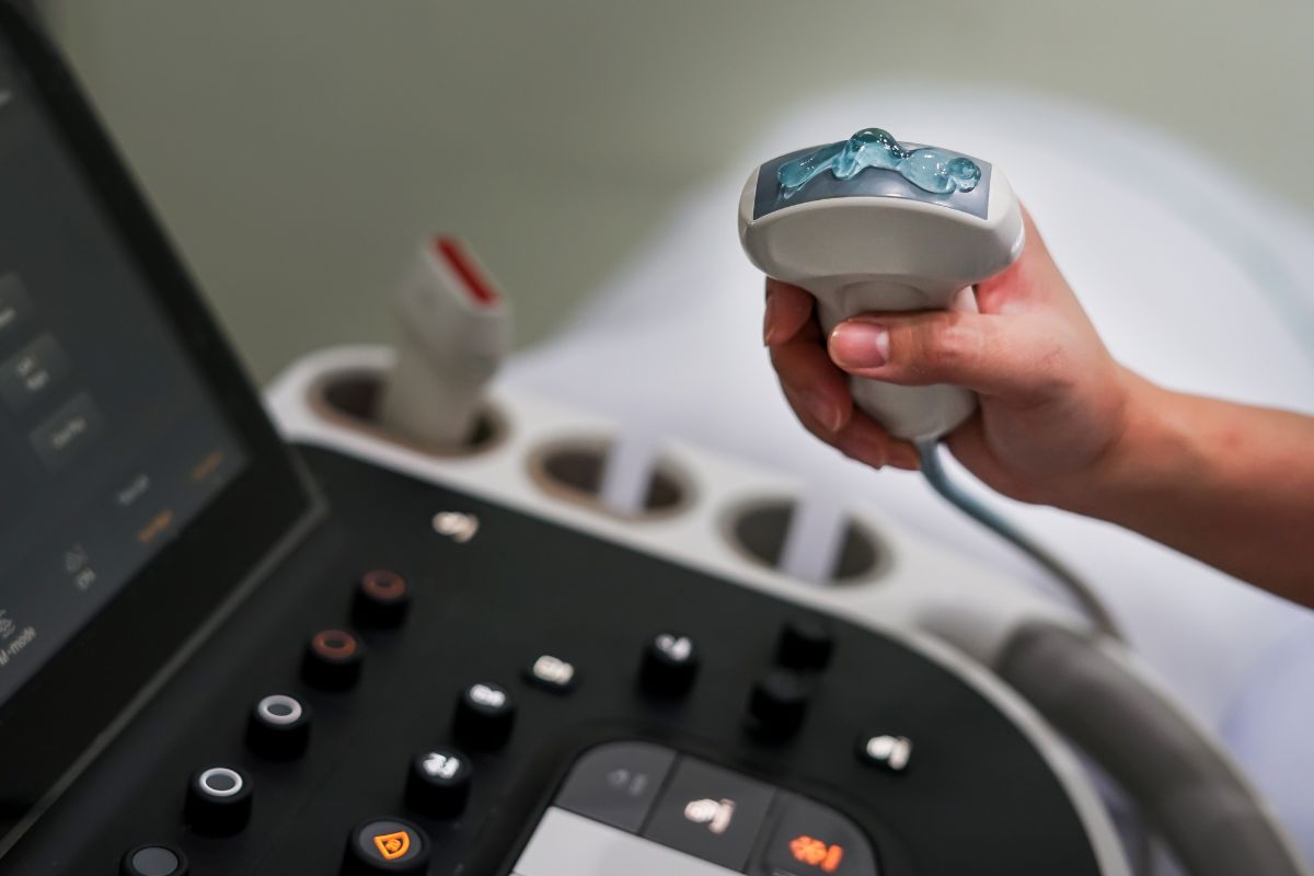
Sound waves that are too high in frequency for the human ear to perceive are ultrasonic. An ultrasound image comes about by injecting a brief burst of sound waves into the patient’s tissues using an ultrasonic transducer (probe). It records echoes when the sound waves go through tissue, and the operator can see them.
This process occurs as the sound waves move through the tissue. Ultrasonography enables the presentation of various photographs and other picture forms. The B-mode image, which illustrates the acoustic impedance of a two-dimensional cross-section of tissue, is the most well-known type of ultrasound picture.
Blood flow and tissue movement over time are further examples of images that might appear. Ultrasonography is different from other imaging techniques because it produces an image in real time.
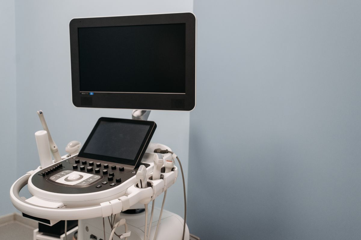
You can use it for imaging at the bedside if it is portable. It is relatively inexpensive and a safe, non-invasive, and useful diagnostic imaging modality with an increasing number of applications. Evaluating horses diagnosed with colic is one of the most prevalent applications of ultrasonography.
Even if its usefulness in the adult horse is in question, ultrasonography is still widely considered irreplaceable in the case of colic in a horse. The size and depth of the abdomen, the partial enclosure of the belly by the ribs, and the degree of gas distension often present are all factors that contribute to the limitations.
Despite these drawbacks, ultrasonography can offer the veterinarian a wealth of information you cannot obtain by any other method. This information can assist the veterinarian in assessing whether or not a horse is suffering from a surgical or medical lesion.
It enables a more accurate interpretation and detection of small intestine strangulating lesions that need surgery and lesions that are not strangulating. It is possible to monitor the progression of small intestine anomalies and collect accurate diagnostic information, leading to earlier surgical intervention.
Ultrasonography is another method to determine the thickness of the intestinal wall characteristic of colitis. The musculoskeletal and respiratory organ systems are two other areas in which ultrasonography can be quite helpful. Through ultrasonography, one can diagnose the severity of tendon injuries and monitor the body’s response to treatment.
Another area in which ultrasonography can be useful is localizing foreign substances. The diagnostic, therapeutic, and prognostic evaluation of pleural or peripheral lung parenchyma problems can all benefit from a sonographic examination of the horse’s respiratory system.
When it comes to detecting effusions, consolidation of the lung, abscesses, and tumors, thoracic ultrasound is more effective than thoracic radiography. Thoracic ultrasonography has proven to be effective in diagnosing foals affected by Rhodococcus pneumonia and determining whether or not they require therapeutic intervention.
One of the drawbacks of thoracic ultrasonography is that a lesion that is deeper within the lung tissue you may not identify if it is underneath normally aerated lung tissue. This is one of the limitations of the test. This is because air and gas reflect the ultrasonic beam, making it impossible to view deeper into the lung tissue.
Unlike adult horses, the size of foals makes them ideal for scanning, overcoming some of the constraints in adult horses. Ultrasonography is advisable in all septicaemic foals and for illnesses such as omphalitis (umbilical infection), which is not always obvious externally, pneumonia, and gastrointestinal disorders.
Most umbilical infections respond to medical intervention alone and can track the response to antibiotic therapy by undertaking recurrent ultrasonographic tests.
In summary, ultrasonography is a very valuable diagnostic modality widely available.
Subtle changes may require more skilled readings but, in general, can give a veterinarian valuable information. You should always evaluate it with the clinical picture of the horse and can complement other diagnostic tests in obtaining a clear diagnosis.


Our Top Equine Vet-Related Books
Atlas of Equine Ultrasonography 1st Edition
By Jessica A. Kidd (Editor), Kristina G. Lu (Editor), Michele L. Frazer (Editor)
The only visual guide to equine ultrasonography based on digital ultrasound technology. Atlas of Equine Ultrasonography provides an equine veterinarian with comprehensive coverage of the horse’s musculoskeletal and non-musculoskeletal areas.
Ideal for practitioners in first opinion or referral practices, each chapter features normal images for anatomical reference followed by abnormal images covering a broad range of recognized pathologies.
The division of the book is into musculoskeletal, reproductive, and internal medicine sections and includes positioning diagrams demonstrating how to capture optimal images. With contributions from experts worldwide, this book is the go-to reference for equine clinical ultrasonography.
Key features include:
- Pictorially based on a wealth of digital ultrasound images covering musculoskeletal and non-musculoskeletal areas and their associated pathologies.
- Each chapter begins with a discussion of normal anatomy and demonstrates how to obtain and interpret the images presented.
- A video library of over 50 ultrasound examinations is available for streaming or download and viewing on the go. The book provides the access details.
You will find the following topics adequately covered in this book:
- Musculoskeletal
- Reproduction
- Internal Medicine
Publisher: Wiley-Blackwell; 1st edition (June 23, 2014)
Language: English
Format: Kindle, hardcover
Hardcover: 520 pages
ISBN-10: 0470658134
ISBN-13: 978-0470658130
Item Weight: 3.71 pounds (1.68 kilograms)
Dimensions: 8.7 x 1.1 x 11.1 inches (22.1 x 2.8 x 28.2 centimeters)
Atlas of Equine Ultrasonography 2nd Edition
By Jessica A. Kidd (Editor), Kristina G. Lu (Editor), Michele L. Frazer (Editor)
About This Book
Ultrasonography is a vital diagnostic tool that you can apply in numerous functions in veterinary practice. In conjunction with relevant clinical information—patient history and physical examination findings, for example—it can be an important aid in the veterinarian’s decision-making process.
Many vets in equine practice rely upon ultrasonography as a mainstay of equine diagnostic imaging on many structures and body systems. Ultrasonography is a useful procedure that is non-invasive and acts in complement to radiography to diagnose the animal’s condition successfully.
This book aims to encourage clinicians to rely further on ultrasonography in their practice. The second edition of Atlas of Equine Ultrasonography provides an updated and expanded revision of the first atlas of ultrasonography in the horse.
The first edition of this important resource was the first pictorially-based book to cover ultrasonography in the horse and remains the only book currently available on the subject.
The current version offers 450 additional images with greater clarity and precision in the images throughout and demonstrates how to obtain images in each body region while offering clinical ultrasonograms that show pathology. Atlas of Equine Ultrasonography readers will also find:
- High-quality clinical ultrasonograms for important musculoskeletal, reproductive, and medical conditions in the horse
- More than 1,500 images, with accompanying concise text describing the images
- A companion website that provides video clips showing dynamic ultrasound exams
Atlas of Equine Ultrasonography is an invaluable reference for any veterinarian evaluating ultrasonograms in equine patients. As a result, this book will be of particular interest to equine specialists, veterinary radiologists, equine practitioners, and veterinary students.
Publisher: Wiley-Blackwell; 2nd edition (November 21, 2022)
Language: English
Format: Kindle, hardcover
Hardcover: 624 pages
ISBN-10: 111951472X
ISBN-13: 978-1119514725
Item Weight: 1.74 pounds (0.79 kilograms)
Equine Diagnostic Ultrasonography 1st Edition
By Norman Rantanen (Author), Angus McKinnon (Author)
About This Book
Ultrasonography is one of the fastest-growing areas in veterinary medicine. This comprehensive and clinically focused resource brings together the leading contributors practicing today under the guidance of Drs. Norman Rantanen and Angus McKinnon.
The book is divided into a section on examination procedures and normal body systems, followed by comprehensive chapters covering all aspects of sonography, including reproductive examination of the mare and stallion, musculoskeletal, cardiovascular, respiratory, abdominal, urinary tract, and neonatal sonography.
It also includes a review of available equipment and different types of ultrasonographic applications. The book covers the following areas at length:
- Principles
- Examination procedures and normal anatomy
- Reproductive ultrasonography – the mare
- Reproductive ultrasonography – the stallion
- Musculoskeletal ultrasonography
- Cardiovascular ultrasonography
- Respiratory system ultrasonography
- Abdominal ultrasonography
- Neonatal ultrasonography
- Miscellaneous applications
Publisher: Wiley; 1st edition (January 15, 1998)
Language: English
Format: Kindle, hardcover
Hardcover: 677 pages
ISBN-10: 0683071238
ISBN-13: 978-0683071238
Item weight: 5.03 pounds (2.28 kilograms)
Dimensions: 8.94 x 1.35 x 11.3 inches (22.71 x 3.43 x 28.7 centimeters)
Equine Reproductive Ultrasonography (Bulletin / Colorado State University, Animal Reproduction Laboratory) Unknown Binding – January 1, 1988
By A. O McKinnon (Author)

About This Book
This text provides an overview of the applications of diagnostic ultrasonography in the horse. It discusses the normal anatomical relationships from an ultrasonographic perspective, the techniques utilized to obtain ultrasonographic images, and the lesions or injuries you can image through ultrasound.
The use of ultrasonographic information to develop a prognosis and follow the case’s progress is also addressed. It covers the more traditional applications: diagnostic ultrasound in theriogenology and flexor tendon and ligament disorders, in evaluating thoracic and abdominal diseases, and Power Doppler to evaluate low-velocity blood flow.
Features the newer applications for musculoskeletal imaging (fractures, osteomyelitis, collateral ligament desmitis, more), theriogenology (ultrasonography of the late-term fetus and intrauterine environment), and spectral and color flow Doppler and their usefulness in the horse.
Explains–step-by-step–how to: perform scans of all body systems, obtain high-quality images, and interpret those images. Discusses how to diagnose subtle lesions: evaluate soft tissue structures of joints, localize fracture fragments, osteomyelitis, and penetrating injuries to synovial structures, follow draining tracks, and locate foreign bodies.
Demonstrates how to use echocardiographic findings to determine the significance of heart murmurs and evaluate myocardial function in horses with poor performance. Shows how to diagnose various abdominal disorders in foals and adult horses.
Presents images of lesions and injuries that you can capture through ultrasound and the techniques used to obtain them. It Includes anatomic drawings to supplement the ultrasonic images. Its content includes the following topics:
- Physics and instrumentation.
- Artifacts and variants.
- Musculoskeletal ultrasonography.
- Thoracic ultrasonography.
- Cardiovascular ultrasonography.
- Abdominal ultrasonography in the adult horse.
- Pediatric abdominal ultrasonography.
- Ultrasonography of the genital tract of the mare.
- Fetal ultrasonography.
- Ultrasonography of the genital tract of the stallion.
- Ultrasonographic evaluation of small parts.
Publisher: Saunders; 1st edition (January 15, 1998)
Language: English
Hardcover: 560 pages
ISBN-10: 0721650236
ISBN-13: 978-0721650234
Item Weight: 4.23 pounds (1.92 kilograms)
Dimensions: 9 x 1.25 x 11.5 inches (22.9 x 3.18 x 29.21 centimeters)
Ultrasonographic Imaging of Equine Pelvis
By Shaaban Gadallah (Author), Ahmed Sharshar (Author), Mohammed El-Sunsafty (Author)

About This Book
Since there is no available data concerning the normal ultrasonographic description of the donkey’s pelvis, although their worldwide distribution, this manual is to provide veterinarians with a detailed comparative ultrasonographic description of the normal anatomy of the pelvis in horses and donkeys.
Pelvis has complex structures, its injuries are common, and its diagnosis using diagnostic means other than ultrasound is difficult. This book aims to make the diagnosis of different pelvic injuries easier.
Publisher: LAP LAMBERT Academic Publishing (April 23, 2019)
Language: English
Paperback: 52 pages
ISBN-10: 6200000417
ISBN-13: 978-6200000415
Item Weight: 3.39 ounces (96.1 grams)
Dimensions: 5.91 x 0.12 x 8.66 inches (15.01 x 0.302 x 21.9 centimeters)
Advances in Diagnostic and Therapeutic Techniques in Equine Reproduction, An Issue of Veterinary Clinics of North America: Equine Practice (The Clinics: Veterinary Medicine Book 32) Kindle Edition
By Marco A. Coutinho da Silva A. Coutinho da Silva (Author) Format: Kindle Edition

About This Book
This issue of veterinary clinics of North America: Equine Practice focuses on advances in diagnostic and therapeutic techniques in Equine reproduction. Article topics include embryo
transfer; breakthroughs in embryo cryopreservation; collection, maturation, and shipment of oocytes for art; icsi, embryo culture, and transfer.
Effects of insulin resistance/Cushing’s on reproduction; use of amh as a diagnostic tool in males and females; endometritis: Managing persistent post-breeding endometritis; endometritis: diagnostic tools for infectious endometritis; endometritis: non-traditional therapies; and more!
eBook Features:
- Highlight, take notes, and search in the book
- In this edition, page numbers are just like the physical edition
- Length: 197 pages
- Enhanced Typesetting: Enabled
- Page flip: Enabled
- Due to its large file size, this book may take longer to download
This book contains several topics that include:
- Embryo Transfer
- Breakthroughs in embryo cryopreservation
- Collection
- Maturation and shipment of oocytes for ART
- ICSI, embryo culture, and transfer
- Effects of insulin resistance/Cushing’s on reproduction
- Use of AMH as a diagnostic tool in males and females
- Endometritis: managing persistent post-breeding endometritis
- Endometritis
- Diagnostic tools for infectious endometritis
- Endometritis
- Non-traditional therapies and more!
ASIN: B01MQEB7QQ
Publisher: Elsevier
Publication date: November 3, 2016
Language: English
File size: 84321 KB
Simultaneous device usage: Up to 4 simultaneous devices, per publisher limits
Text-to-Speech: Enabled
Screen Reader: Supported
Enhanced typesetting: Enabled
X-Ray: Not Enabled
Word Wise: Not Enabled
Print length: 197 pages
Page numbers source ISBN: 0323477542
Lending: Not Enabled
FAQ on Equine Ultrasonography the Books Addresses
What is Ultrasound Used for in Horses?
As an equestrian veterinarian, you can obtain images of the orthopedic soft tissues in horses, such as tendons and ligaments, through equine veterinary services. You can use ultrasonography to examine smaller and major joints, such as the stifle. In addition, we utilize ultrasonic imaging when researching wounds and even when looking for fractures in the pelvis.
Which Probe for Which Application?
In general, there are distinct probes designed specifically for each application; nevertheless, there are some instances in which these probes are interchangeable. They include
- The micro-convex: This probe is the superior option for doing abdominal ultrasounds on tiny animals. Its narrow head is ideal for squeezing into the abdominal cavity, and the expansive sector provides an excellent view of the organs found within the body.
- Phased Arrays / Sector: These are special cardiac probes known as phased and sector arrays. Their development is specifically for cardiac imaging, and as a result, their contact point is quite small. This makes it easier to see clearly through the ribcage. They can also produce larger frame rates and better detect blood flows moving at higher velocities.
- The convex and curvilinear: These probes often have lower frequencies and are for deeper scanning; as a result, they are more appropriate for use with equine patients. The larger probe head makes it more difficult to keep contact up and under the ribs and causes the smaller animals to experience significant discomfort.
- Linear: These are for imaging at a more superficial level, and their frequencies and contact areas tend to be greater. They also have a bigger surface area, which provides better near-field resolution. They are most effective when scanning objects with a diameter of less than (1.97 to 2.4 inches) 5 to 6 centimeters, such as cat abdomens, intestines, spleens, kidneys, etc.
You can perform musculoskeletal imaging with linear probes that operate at higher frequencies.
Several applications for the probes go beyond their “typical” function, and these applications are available. It is possible to examine the heart with a micro-convex, but the results will not be completely accurate.
Because of the shape of the micro convex head, you will experience a significant amount of rib shadowing, and the Doppler performance will not compare favorably to that of specialized cardiac probes.
The same is true for phased array probes; although you can use them for abdominal scanning, these probes are not ideal because of their limited close field of vision, making superficial scanning structures difficult. Nonetheless, you can use these probes.
How Do You Explain Ultrasonolgy Pictures To Your Client?
As horse vets, you now have the knowledge to comprehend the images that appear on an ultrasound machine; however, your equine patient can find them difficult to understand.
It’s possible that they don’t even realize what ultrasound imaging is, what it can perform, or how useful it may be as a diagnostic tool. While describing horse owners to ultrasound pictures, the following are some pointers to keep in mind.
Tell Your Client What Ultrasound is and How It Works
In a nutshell, ultrasound machines create images by employing sound waves. Therefore, you should explain to your client that the technology uses painless, noninvasive, high-frequency sound waves that are inaudible to the human ear and completely safe to expose their horse to.
Assure your customer that the sound does not induce an uncomfortable sensation as it travels through their horse’s body; however, it produces echoes that determine the distance between internal organs and tissues and their size and shape.
Explain Why Ultrasound Imaging is a Good Diagnostic Tool
Your customer may be aware that it is “standard procedure” to perform an ultrasound on a pregnant mare; nevertheless, he may not grasp the rationale for this practice. Inform him that ultrasound scans are the most reliable method for determining whether or not his mare is pregnant and whether her fetus is developing normally.
You can also let your client know that ultrasound machines disclose potentially harmful circumstances that you can’t identify with touch alone, such as many pregnancies or anomalies in the uterus.
Black and White Sheds Light
Your customer, who see the world in technicolor, may take aback when they realize that ultrasound images are in shades of black, white, and gray. You are at ease with this monochrome image, but the average horse owner might be wary of modern technology that does not use full, vivid color!
Tell him that the only things that are truly important to view on the monitor are light and dark and that this is because the strength of the returning signal translates into a level of brightness displayed on the screen.
The image gets brighter as the density of the tissue increases; hence, the fluid appears black, dense solids like bladder stones appear dazzling white, and the interior organs and structures will appear in shades of grey. Your client will easily understand what the ultrasound image implies after they have this foundational knowledge.
Use the Fruit Analysis to Explain the Orientation
Oranges are a fantastic visual aid for demonstrating how the transducer works in an ultrasound machine. Ask your customer to imagine an orange sliced vertically from the stem to the navel and horizontally across the diameter. Depending on the direction you cut the rings and segments, they each have a unique appearance.
Make use of this visualization method to assist your client in comprehending that by adjusting the position of the ultrasound probe, you can visibly slice the internal organs in whichever direction is necessary to receive the information you are looking for.
Keep it Simple
A significant number of horse owners have a deep interest in equine physiology, and it is safe to say that the number of those horse owners who breed their mares is significantly higher. But don’t confuse that fascination with a profound understanding of veterinary practices and procedures with becoming a vet for horses.
You know how organisms operate, but you must be careful not to confuse your customer by providing too many specifics. Instead, draw attention to fundamental features that are easily identifiable, such as the beating heart of a fetus, the distinctive form of a kidney, or the specks of pus floating around in an abscess.
Create a Keepsake
There are a significant number of horse owners who have a profound interest in equine physiology, and it is safe to say that the number of those horse owners who breed their mares is significantly higher than the number of those horse owners who have an interest in equine physiology and have an interest in breeding their mares is significant.
However, they should not confuse this fascination with an in-depth knowledge of the processes and practices used in equine veterinary care. You understand how living things function, but you must be careful not to baffle your client by presenting an excessive amount of specifics in your explanations.
Instead, focus on essential characteristics that are straightforward to recognize, such as the beating heart of a fetus, the particular shape of a kidney, or the flecks of pus floating around in an abscess.

That’s a Wrap
When it comes to Equine Ultrasonography, there is a lot you need to understand as an equine veterinary technician. The good news is that there are very many books and equine veterinary journal you can read about Equine Ultrasonagraphy. However, Atlas of Equine Ultrasonography 1st Edition stands out from the rest.
It has several chapters that touch on the key ultrasonagraphy that gives you a detailed explanation of equine vet that you need to know. It is a must-have book, and you will not regret having it in your collection of books.